Pentax 2013 Annual Report Download - page 46
Download and view the complete annual report
Please find page 46 of the 2013 Pentax annual report below. You can navigate through the pages in the report by either clicking on the pages listed below, or by using the keyword search tool below to find specific information within the annual report.-
 1
1 -
2
-
3
-
4
-
5
-
6
-
7
-
8
-
9
-
10
-
11
-
12
-
13
-
14
-
15
-
16
-
17
-
18
-
19
-
20
-
21
-
22
-
23
-
24
-
25
-
26
-
27
-
28
-
29
-
30
-
31
-
32
-
33
-
34
-
35
-
 36
36 -
 37
37 -
 38
38 -
 39
39 -
 40
40 -
 41
41 -
 42
42 -
 43
43 -
 44
44 -
 45
45 -
 46
46 -
 47
47 -
 48
48 -
 49
49 -
 50
50 -
 51
51 -
 52
52 -
 53
53 -
 54
54 -
 55
55 -
 56
56 -
57
-
58
-
59
-
60
-
61
-
62
-
63
-
64
-
65
-
66
-
67
-
68
-
69
-
70
-
71
-
72
-
73
-
74
-
75
-
76
-
77
-
78
-
79
-
80
-
81
-
82
 |
 |

Business Overview
Copyright 2013 © HOYA CORPORATION
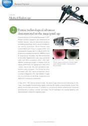
2Pentax technological advances
demonstrated in the mega pixel age
Clinical images provided by Dr. Shinya Odajima
and Dr. Mitsuhiro Fujishiro, the University of
Tokyo Hospital.
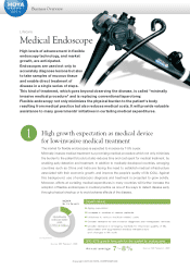
Medical Endoscope
Lifecare
Since the launch of a bronchial fiberscope in 1977,
Pentax has been engaged in the development of
medical devices, using its optical and precision
processing technology, which was accumulated in
i t s c a m e r a b u s i n e s s . W h i l e P e n t a x w a s
consolidated with Hoya in August 2007 and
subsequently merged with it in March 2008, the
endoscope business has kept the PENTAX brand
for its products. At present, we offer endoscopes
for bronchial, digestive tract, ears, nose, and throat
uses, and video processors which offer high-
definition endoscope images. In October 2012, we
released EPK-i7000, the highest-end model of the
i-series, that is also the next-generation high-
function video processor. Combining the video
processor with the i-series endoscope, which
mounted a Megapixel CCD, high-definition images
can be provided to facilitate diagnosis and
treatment in endoscopic examination.
In May 2013, "OE" (Optical Enhancement), the latest image enhancement technology for the
i-scan, was released.This technology digitally enhances the visibility of blood vessels, ducts of the
glands and surface structures, in addition to conventional optical enhancement functions
(enhancement of surface, contrast, and tones.) The OE facilitates more precise detection and
characterization of lesions in digestive organ.